|
Sternocostal surface
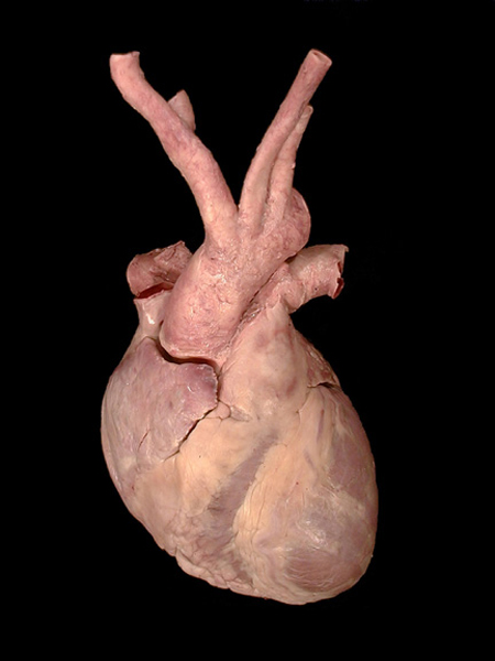
The heart removed from the cavity. The parietal pericardium is also removed. (The visceral is adherent to the heart and forms the epicardium.) Note the right atrium, right auricle, right ventricle and the left ventricle. The vessels seen are the right pulmonary artery, SVC, aorta with its branches and pulmonary trunk leading to the left pulmonary artery. There are two bands of fat covering the atrio-ventricular and anterior inter-ventricular grooves.
Dr. S. Chandra
Dr. S. Chandra, Faculty of Medicine, Memorial University of Newfoundland
| |